UV fluorescence and UV transmittance


In the 300 - 400 nm spectral range, fluorescence in proteins originates predominantly from tryptophan (Trp) and tyrosine (Tyr) residues. By choosing an exciting wavelength that induces fluorescence from tryptophan and, to some extent, tyrosine, most protein molecules can be detected.
Fluorescence from Trp dominates over fluorescence from Tyr. Trp may also quench Tyr fluorescence. Trp fluorescence itself may be quenched by the presence of nearby charged residues, bound ligands, and cofactors, due to energy or electron transfer. The fluorescence quantum yield of Trip in proteins varies from 0 to 40%.
For example, hen egg white lysozyme, with 3 Tyr residues and 6 Trp residues, has a near-UV fluorescence quantum yield of 12%, owing mainly to two Trp residues that trap the excitation energy.
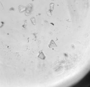
Left Top: Fluorescence from protein crystals buried in precipitate;
Left Bottom: Fluorescence from protein crystals lacking Trp


Prolonged excitation of Trp residues can result in photochemical conversion of amino acid and lead to a loss of near-UV fluorescence. Repeated high-intensity excitation at shorter wavelengths can cause denaturing and/or degradation of the protein. UVEX imager employs efficient optics and camera to allow operation at low excitation intensities.
UV transmission is a more dependable means of identifying the protein crystal when UV fluorescence is quenched by small molecules bound to the protein.
When the apo-protein is fluorescent and the complex is not, comparison of UV fluorescence image with UV transmission image provides a powerful method for detecting crystals of the complex.
transmission imaging also is very suitable for crystals of nucleic acids and does not require staining the crystals with fluorescent dye for identification.
The optics and the camera of UVEX are suited for UV transmission imaging. UV transmissions requires UV-transparent optics, filters different than needed for UV fluorescence, and a camera sensitive down to 280 nm, criteria that are met by UVEX imagers.
Right: Images of crystals of DNA-protein complex -
Top: Transmission
Middle: UV fluorescence
Bottom: UV transmittance / absorbance
Publications where UVEX imager has had significant mention
-
From Screen to Structure with a Harvestable Microfluidic Device, Stojanoff et al. (2011), Acta Crystallogr F 67(Pt 8) 971-5
-
Use of Transmission Electron Microscopy to Identify Nanocrystals of Challenging Protein Targets, Stevenson et al. (2014) Proc Natl Acad Sci USA 111 (23): 8470-5
-
Transmission Electron Microscopy for the Evaluation and Optimization of Crystal Growth, Stevenson et al. (2016) Acta Crystallogr D 72(Pt 5) 603-15
-
Molecular mimicry in deoxy-nucleotide catalysis: the structure of dGTPase reveals the molecular basis of dGTP selectivity, Barnes et al. (2019) Proc Natl Acad Sci USA, 116 (19) 9333-39
-
Hallucinating symmetric protein assemblies, Wicky, et al. (2022) Science, 378 (6615) 56-61
-
Design of stimulus-responsive two-state hinge proteins, Praetorius, et al. (2023) Science, 381 (6659) 754-760
-
Hallucination of closed repeat proteins containing central pockets, An, et al. (2023) Nature Structural & Molecular Biology https://doi.org/10.1038/s41594-023-01112-6
-
De novo design of highly selective miniprotein inhibitors of integrins αvβ6 and αvβ8, Roy, et al. (2023) Nature Communications https://doi.org/10.1038/s41467-023-41272-z
-
Directing polymorph specific calcium carbonate formation with de novo protein templates, Davila-Hernandez, et al. (2023)Nature Communications 14, 8191.
-
Precisely patterned nanofibres made from extendable protein multiplexes, Bethel, et al. (2023) Nature Chemistry 15 1664-71.